


The Modular Organization of Pain Brain Networks: An fMRI Graph Analysis Informed by Intracranial EEG
Camille Fauchon, David Meunier, Isabelle Faillenot, Florence B. Pomares, Hélène Bastuji, Luis Garcia-Larrea and Roland Peyron (NEUROPAIN CRNL)
Cerebral Cortex Communications, 2020, 1–13
Abstract
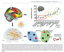
Intracranial EEG (iEEG) studies have suggested that the conscious perception of pain builds up from successive contributions of brain networks in less than 1 s. However, the functional organization of cortico-subcortical connections at the multisecond time scale, and its accordance with iEEG models, remains unknown. Here, we used graph theory with modular analysis of fMRI data from 60 healthy participants experiencing noxious heat stimuli, of whom 36 also received audio stimulation. Brain connectivity during pain was organized in four modules matching those identified through iEEG, namely: 1) sensorimotor (SM), 2) medial fronto-cingulo-parietal (default mode-like), 3) posterior parietal-latero-frontal (central executive-like), and 4) amygdalo-hippocampal (limbic). Intrinsic overlaps existed between the pain and audio conditions in high-order areas. Neocortical modules were interrelated via “connector hubs” in dorsolateral frontal, posterior parietal, and anterior insular cortices, the antero-insular connector being most predominant during pain. These findings provide a mechanistic picture of the brain networks architecture and support fractal-like similarities between the micro-and macrotemporal dynamics associated with pain. The anterior insula appears to play an essential role in information integration, possibly by determining priorities for the processing of information and subsequent entrance into other points of the brain connectome.