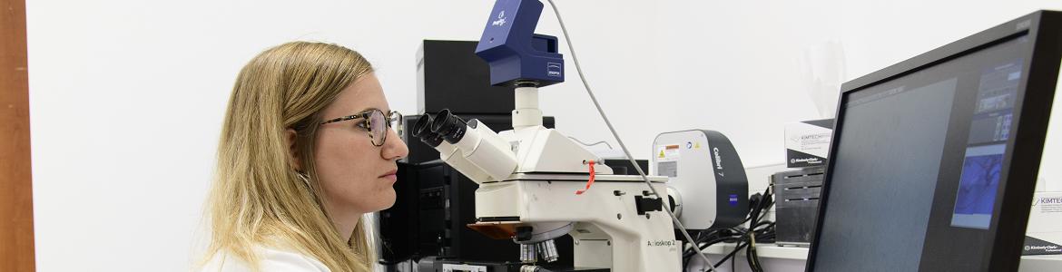
Skills
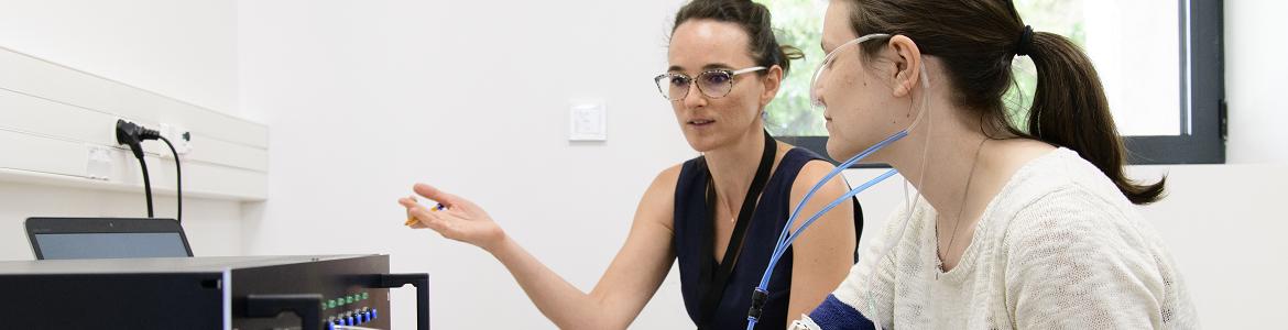
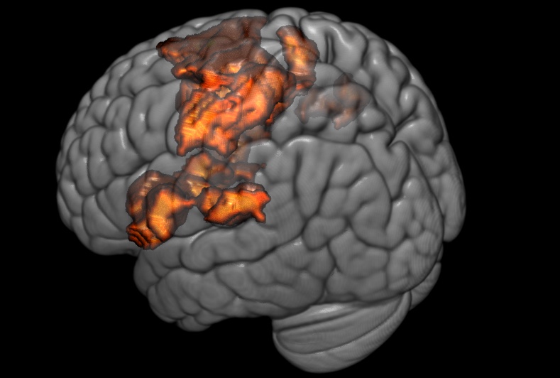 activations during nociceptive stimulations
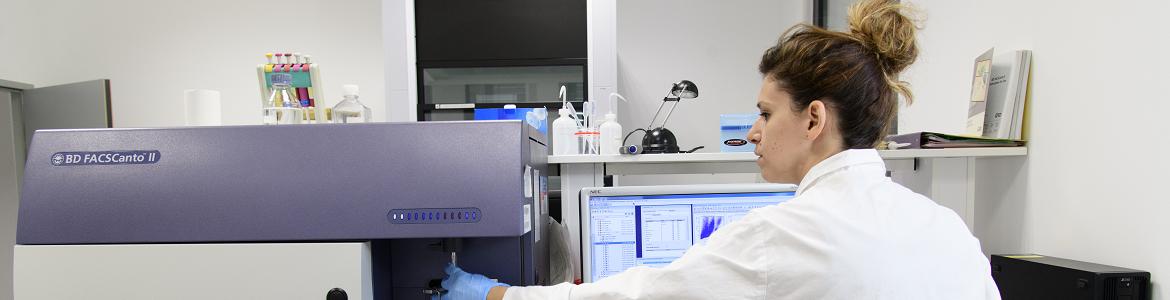
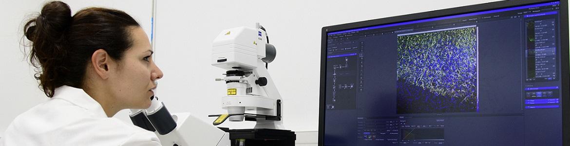
- Image analysis: Functional activation or connectivity with MRI, perfusion with ASL
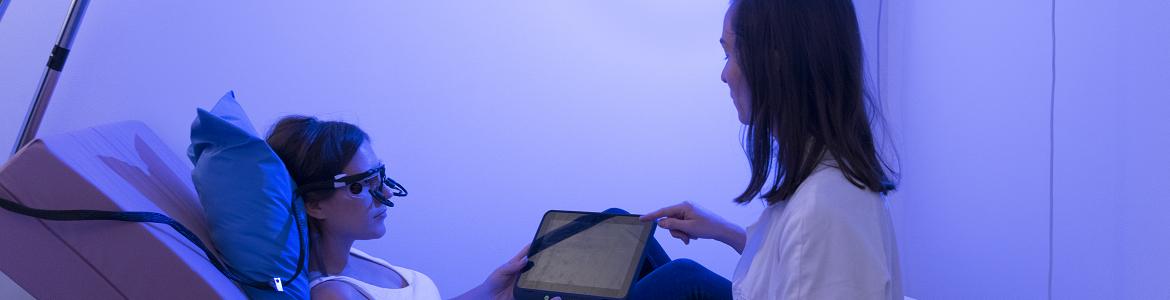
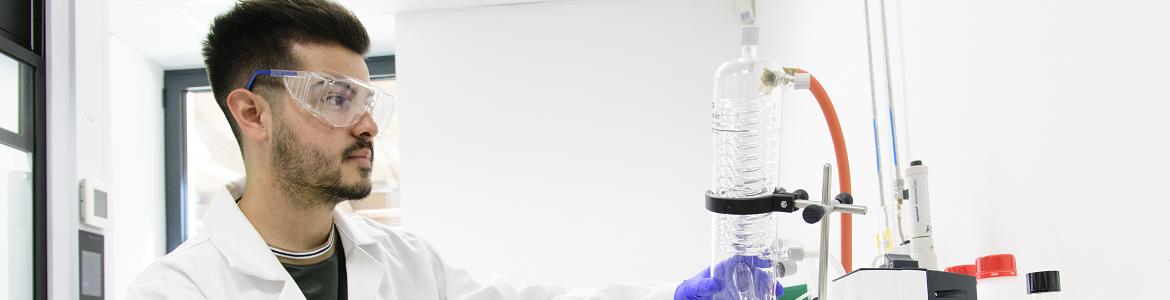
- Preparation of MRI experiments
- Anatomical location and on use of human atlases.
Developed tools
- Macroanatomical and probabilistic atlas of human insula
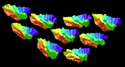 Macroanatomical subdivisions of 9 insulae
I segmented the insular of 30 subjects.
Theses segmentations were included in the human atlas of A. Hammers et al. which consisted in 95 regions (Faillenot et al, NeuroImage 17).
These regions are also available in MNI space.
- DLPFC localizations according to different methods for rTMS stimulation
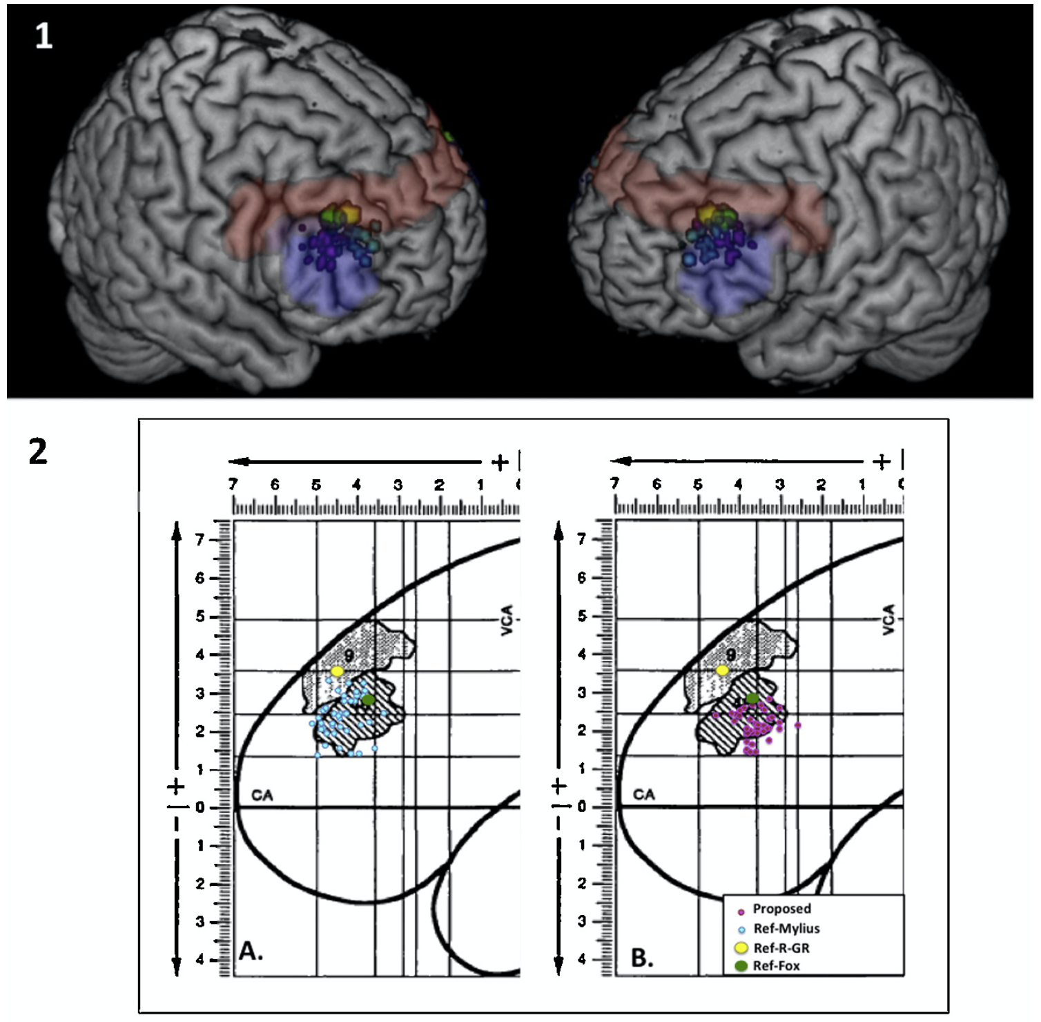 DLPFC location
SPM batches to include white voxels in the T1 anatomical MRI, for example to make a rTMS targeting. The voxels chosen, the rTMS target, should be inside the MNI space. The batch first compute parameters to convert the MNI space towards the subject space and then apply these parameters to the target image. Batches are provided with some MNI image of the SII, DLPFC, posterior and anterior insula (Pommier et al, 17). Ask me the batches or dowload from the GitHub of our lab.
Selected papers
Investigating brain dysfunction in neuropathic pain with MRI. Roland Peyron, Siloé Corvin, Camille Fauchon, Isabelle Faillenot. Brain Communications, 2025, 7 (3), pp.fcaf196.
Macroanatomy and 3D probabilistic atlas of the human insula. Faillenot I, Heckemann RA, Frot M, Hammers A. Neuroimage. 2017 Apr 15;150:88-98.
And the last ones:
Parenting and empathy capabilities drive brain response to pain cues in baby cries - Camille Fauchon; Siloé Corvin; Isabelle Faillenot; Hugues Patural; David Reby; Roland Peyron; Nicolas Mathevon ID: PAIN-D-25-00902R2
Cardiac autonomic responses to cortical electrical stimulation: a SEEG study. Chouchou F, Soulier H, Pichot V, Mauguière F, Faillenot I, Guénot M, Hermier M, Jung J, Montavont A, Catenoix H, Isnard J, Roche F, Rheims S, Mazzola L.Neuroimage. 2025 Sep;318:121423. doi: 10.1016/j.neuroimage.2025.121423. Epub 2025 Aug 15.PMID: 40819828
list in pubmed
papers in open access