NEUROPAIN

Central Integration of Pain in Human
Objectives

NEUROPAIN develops physiological, pre-clinical and clinical research on pain. It develops its research working

almost exclusively on human beings

The group is committed with three main research axes:

  1. the study of cortical mechanisms leading to, and modulating pain perception in humans (Pain Physiology);
  2. the use of neurophysiological techniques to improve our understanding, diagnosis and prediction of neuropathic pain (PathoPhysiology of Pain);
  3. the improvement of cortical neurostimulation procedures for pain relief through a better comprehension of their mechanisms (NeuroStimulation). 
Equipments

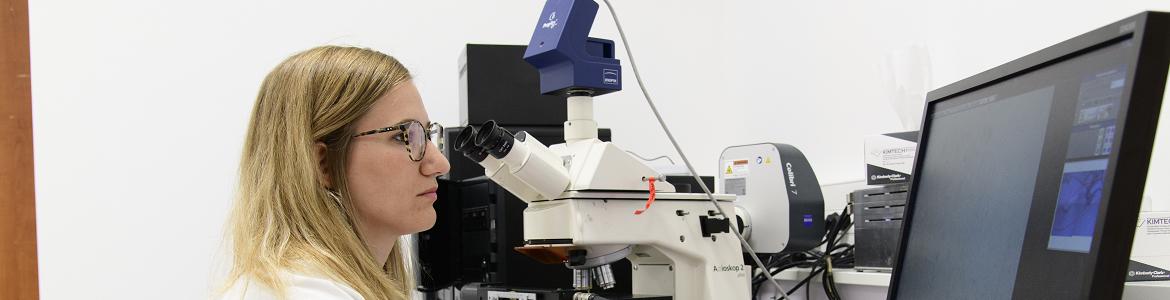
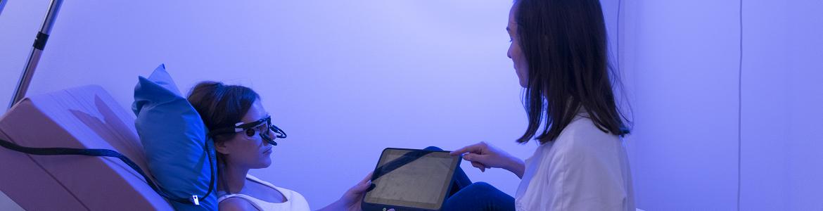
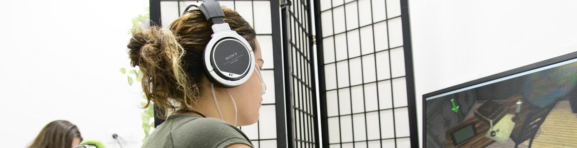
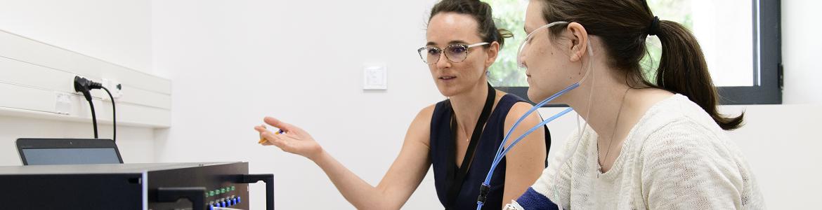
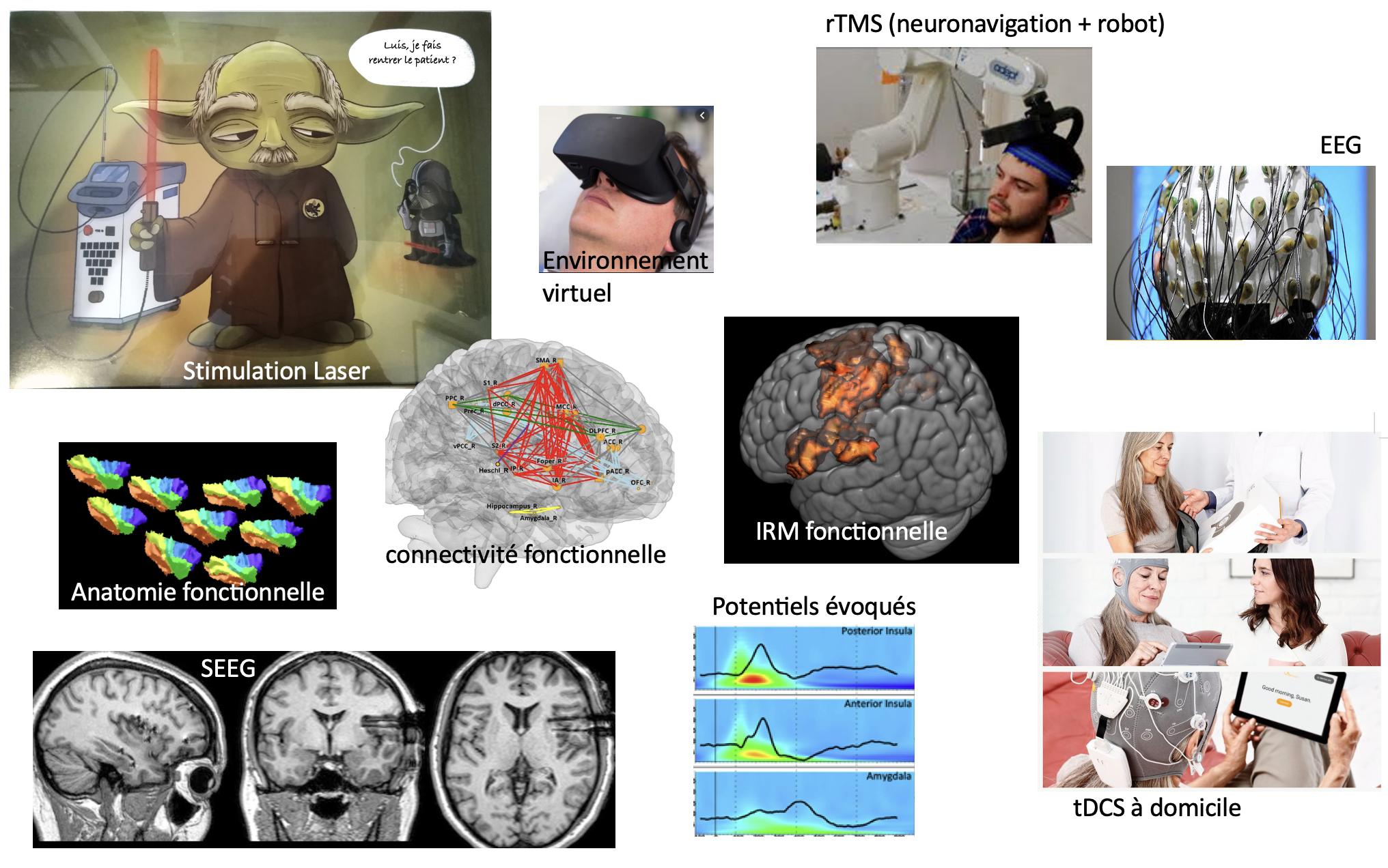
The team is physically located within the Neurological Hospital of Lyon and the University hospital of Saint-Etienne. Most of the team members have clinical activities, hence ensuring direct access to clinical facilities and patient wards. NeuroPain has dedicated systems for high-density electrophysiology (2x128- channel EEG) and cortical stimulation (rTMS, tDCS, epidural and intracortical stimulation), as well as 3D neuronavigation facilities with robotized arm and several multi-modal sensory stimulation systems, including two nociceptive-specific Nd:YAP laser stimulators. We have access to MEG, fMRI and PET- scan facilities, as well as to the platform for intracranial EEG, which is coordinated by one of our team members. NeuroPain is also founding member of, and directly involved in the development of the platform “Neuro-Immersion”, coupling Virtual Reality with HD-EEG, TMS and 3-D real-time kinematics. 


Human resources

NeuroPain Team 21
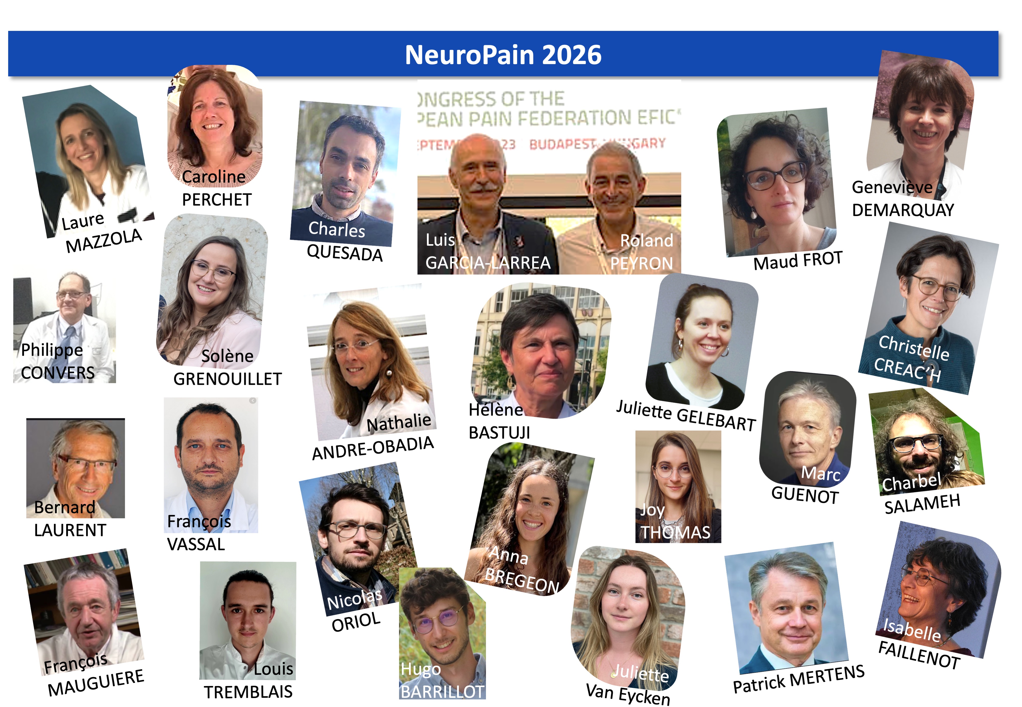
NeuroPain joins efforts of 20 tenured members: neurobiologists, neurologists, psychologists, neurosurgeons, research engineers.

  • 3 researchers  INSERM or University Lyon1
  • 3 technical / ingeneer support staff 
  • 13 clinicians   (including 2 with 50% secured research time)

equipe22

Methods and Techniques

Psychophysics, EEG, intracranial EEG, anatomical and functional MRI, PET, neuronavigated rTMS, tDCS

Signal and image processing, brain oscillations, source modeling, connectivity.equipement

Research Project

Axis 1. Cortical physiology: the construction of pain

Axis 1.1. Functional characterisation of the insular lobe in humans

The human posterior insula is a multisensory area containing networks responsible for the initial steps of pain processing. The posterior insula and adjacent medial operculum are the major projection sites of the spinothalamic system in primates, the only cortical areas where focal stimulation induces acute pain, and where focal lesions selectively interfere with nociception. Our work has underscored the insula as an entry point into complex networks leading to conscious pain (Garcia-Larrea & Bastuji, PNBP 2018; Garcia-Larrea & Mauguière, Handbook Clin Neurol 2018). 

Isabelle Faillenot and Maud Frot (NeuroImage, 2017) developed a tri-dimensional computerised probabilistic atlas of the human insula and created probability maps in the MNI152 stereotaxic space. 

Alexandra Montavont and colleagues analysed intracranial EEG ictal recordings and  data from intracranial stimulation. The posterior insula and adjacent operculum, but not SI nor the cingulate region, were systematically involved at the onset of seizures.

The multisensory nature of the posterior insular region was studied by Laure Mazzola, who characterised the different sensory percepts evoked by electrical stimulation of this area. 

(iv) Functional segregation of posterior and anterior insulae. Maud Frot investigated the rapid posterior-to-anterior information flow in the insula, and showed a 40-70 ms time lag between posterodorsal and anteroventral insular sectors (Frot et al, Hum Brain Mapp, 2014). The anterior insular sectors do not behave as sensory cortices but are rather involved in the cognitive and emotional appraisal of sensory stimuli. Hélène Bastuji assessed the interactions between the insula and the limbic system by studying functional correlation between the posterior and anterior insulae, and the amygdalar nucleus.

Our results support a model whereby parallel activation of sensory and limbic nociceptive networks (from spino-thalamic and spino-amygdalar routes) converge onto the anterior insula. This region may integrate limbic with sensory input, explaining why it has been considered the initiator of decisions on ‘painfulness’. Insular-amygdalar connections might also contribute to explain the changes in subjective time estimation that occur while suffering pain (Rey et al, Sci Rep, 2017).

Axis 1.2. From nociception to conscious pain

Hélène Bastuji and colleagues studied human intra-thalamic recordings in 3 nuclei receiving spinothalamic (STT) projections. This study indicates that a single nociceptive volley is able to trigger the sensory, cognitive and emotional activities underlying complex pain experiences (Bastuji et al, Cereb Cortex, 2016). 

Different cortical activation phases were disclosed according to their relation with conscious pain perception. Pre-conscious activity was recorded in posterior insula, mid-cingulate and amygdala, while voluntary reactions signalling perception coincided with activity in anterior insula, prefrontal and posterior parietal areas (Bastuji et al, Hum Brain Mapp, 2016; Bradley et al, Hum Brain Mapp, 2017). The relevance of parieto-frontal activity to stimulus awareness was also supported by studies of Mazza et al (2014) and Peter-Derex et al (2015).

This series of studies support the notion that the aversive experience we call “pain” results from the coordinated activation of multiple brain areas. The so-called “pain matrix” appears not as a fixed arrangement of structures but rather as a fluid system of interacting sub-networks, beginning in parallel within sensory, motor and limbic areas and expanding to higher-order networks supporting salience, executive control and the passage from pre-conscious nociception to  conscious  pain. Our continuous work in this domain led to the Edition and coordination of the multi-authored book “Pain and the Conscious Brain” (Garcia-Larrea & Jackson Eds, 2016).

Axis 1.3. Cognitive & emotional modulation of pain responses

Our pain and the pain of others. 

Ten years ago we described the phenomenon of ‘compassionate hyperalgesia’, whereby observing pain in others enhances our own pain. Under the supervision by Maud Frot, Claire Czékala (J Pain, 2015) compared the time needed to discriminate the expression of pain in a human face and Anaïs Chapon (Sci Rep 2019) showed that the mere observation of painful body contacts elicited hyperalgesia and enhanced vegetative reactions in the observer.

Reverse empathy 

It refers to how our pain is influenced by the (un)empathetic comments from others. Roland Peyron and Camille Fauchon developed an ‘ecological experiment’ where caregivers talk about the subjects’ pain, as in hospital rounds. Subjective pain was significantly attenuated by empathetic comments, while unempathetic remarks, although accelerating heart rate, failed to modulate pain (Fauchon et al, Eur J Neurosci, 2017; Auton Neurosci, 2018).

Axis 1.4. Sleep modulation of pain responses

Léa Claude and Hélène Bastuji (J Physiol, 2015), showed that sleep spindles do not block nociceptive responses, and may even enhance cortical potentials to noxious laser during sleep. In parallel, continuous collaboration with Eric Halgren’s group (Univ California San Diego) has shown that spindles do not precede nor accompany periods of decreased cortical activity (“downstates”, or periods when most cortical cells stop firing) but rather follow such states (Gonzalez et al, J Neurosci, 2018). Spindles might therefore signal the end of a period of decreased activity, and hence reflect active procedures, including memory encoding. In accordance, cortical downstates might be caused by cortically-induced disruption of spindling (Mak-McCully et al, PlOs Comput Biol, 2014; Nat Commun 2017). Consistent with these data, very recent results from the lab show that nociceptive-evoked-potentials are maximal when the stimulus is delivered just before, or during a spindle, as compared with the periods following it (Bastuji et al, in preparation).

Axis 2. From physiology to clinical diagnosis: Clinical & translational studies

Axis 2.1. Predicting central pain

Thalamic pain (Dejerine-Roussy’s syndrome) is a severe and treatment-resistant type of central pain. While lesions within the ventrocaudal thalamus carry the highest risk to generate pain, its emergence in individual patients remains impossible to predict. Vartiainen et al (Brain, 2016) showed that lesion of spinothalamic afferents to the posterior thalamus appears determinant to the development of central pain after thalamic stroke. Sorting out of patients at different risks of developing thalamic pain may be achievable at the individual level using easy-to-develop methods of lesions localization and spinothalamic evoked-potentials.

Axis 2.2. Detecting poorly known and mis-diagnosed pain syndromes

While differential diagnosis of neuropathic pain (NP) has greatly improved in recent years, many patients with NP still remain misdiagnosed because of little awareness of ‘hidden NP syndromes’. Our team has reported on the detection of two such neuropathic pain conditions, as well as that of uncommon painful seizures.

Nathalie André-Obadia and their colleagues studied sensory conduction, sympathetic responses and intra-epidermal fibre density in Morvan syndrome patients, and showed that pain was neuropathic, related to selective involvement of small nerve fibres (Laurencin et al, Neurology, 2015).

Philippe Convers et al (Eur J Pain 2020) not only demonstrate that central post-stroke pain can be secondary to small peduncular lesions, but also shows that the condition can be misdiagnosed as simulation or malingering for years.

Alexandra Montavont et al (Neurology, 2015) identified the posterior insula and adjacent operculum as systematically involved at the onset of painful somatosensory seizures. Stimulation of the posterior insula reproduced painful somatosensory symptoms of same quality as those occurring during seizures. 

Axis 2.3 Stimulating pain fibres without pain: intraepidermal electrode

Intra-epidermal electrical stimulation (IES) activates selectively Aδ fibers subserving spinothalamic-mediated sensations (“pricking”) at non-painful intensities, and hence might be useful to replace laser or thermode pulses that some patients dislike. Koichi Hagiwara (Eur J Neurosci, 2018) recorded intracortical responses in 11 patients with stereotactically implanted electrodes in a variety of cortical areas. Intraepidermic stimulation can activate the STT system at low energy levels, with little recruitment of affective-motivational networks and may be of clinical use for the diagnosis of neuropathic pain.   

Axis 3. Neurostimulation for pain relief: from knowledge to cure

Axis 3.1. Predicting the effect of cortical stimulation using non-invasive procedures

Clinical efficacy of neurosurgical motor cortex stimulation (MCS) has been reported in ~50% of patients with drug-resistant neuropathic pain, and repetitive magnetic cortical stimulation (rTMS) is considered a predictor of MCS effect. We assessed the long-term pain relief (2 to 9 years) after epidural MCS and its pre-operative prediction by rTMS. The results provide the longest follow-up of MCS so far; they indicate that half of the patients may retain a significant benefit after 2–9 years of MCS, and that this effect can be reasonably predicted by preoperative rTMS (André-Obadia et al, Pain Physician, 2014, Pommier et al, J Neurosurg, 2018).

Axis 3.2. Mechanisms of motor cortex stimulation: Human and animal models

Single unit recordings during MCS in cats. Our group was pioneer in proposing mechanistic models of MCS action based on human PET-scan studies. An essential step of these models was a cortico-thalamic initial activation. In collaboration with Sandra Kobaiter and Joseph Maarrawi (Pain, 2018), we investigated the effects of MCS on single-unit activities of the thalamic VPL nucleus in cats. 

Somatotopic or not? Relations between cortical stimulation and pain localization.

The influence of somatotopic matching between pain topography and cortical stimulation sites might differ in rTMS and MCS and remains controversial. André-Obadia et al (Eur J Pain, 2017) found that rTMS over the hand motor area was significantly superior to face rTMS, and this whatever the localisation of the pain.

Axis 3.3. Cortical stimulation for pain relief: the insular connection

About 50% of patients fail to respond with adequate pain relief to motor cortex stimulation. This has led to the quest of new targets, one of the best candidates being the posterior insula. In collaboration with the Federal University of Sao Paulo (Brazil) we have developed new procedures for insular neuromodulation, tested in animal models, and being now translated into clinical practice.   

Targeting the insular cortex for pain relief: animal models and clinical translation

The behavioural effects of electrical stimulation of the posterior insular cortex were tested in an experimental rat model of peripheral neuropathy (Dimov et al, Behav Brain Res, 2018) reinforced the potential interest of insular stimulation in neuropathic pain, via modulation of opioid /cannabinoid systems.

We collaborated with Daniel Ciampi de Andrade (University of Sao Paulo) in a clinical trial using non-invasive stimulation of the posterior insula in 98 patients with central drug-resistant neuropathic pain. After 12 weeks of stimulation, although insular stimulation did increase heat pain thresholds in patients, it failed to modify significantly clinical pain or pain interference with daily life (Galhardoni et al, Neurology, 2019). 

Reaching the insula through the back door: vestibulo-insular stimulation

Vestibular afferents converge with nociceptive afferents within the posterior insula, and vestibular sensations can be elicited by electrical stimulation of the postero-insular cortex (Mazzola et al, Annals of Neurol, 2015); vestibular input can modulate nociception, and caloric vestibular stimulation has been shown to reduce experimental and clinical pain. Since caloric stimulation is unpleasant, we developed non-invasive vestibular activation using bi-mastoid galvanic stimulation (GVS) and assessed its effects on experimental pain (Hagiwara et al, Brain Stim 2019). GVS appears as a potentially useful, well-tolerated procedure for the relief of pain, probably through physiological interaction within insular nociceptive networks.

fMRI during insular or motor tDCS

Transcranial direct-current stimulation (tDCS) is emerging as a possible alternative to rTMS procedures for pain relief, and the large distribution of the induced currents might render easier the activation of deep structures. Claire Bradley and Isabelle Faillenot used fMRI to assess regional brain perfusion during two modes of tDCS centred over the motor areas, or the operculo-insular cortex (Bradley et al, submitted). Brain perfusion changes from operculo-insular and motor cortex tDCS converged in multiple brain areas. Since both motor and operculo-insular montages induced changes in posterior insula, it follows that conventional (motor) tDCS might be sufficient to ensure modulation of the insular cortex in patients.

Axis 3.4. Optimising non-invasive stimulation in real life

The routine clinical use of non-invasive neuromodulation is hampered by the limited duration of clinical benefits. Patients responding to non-invasive procedures are confronted with the dilemma of either going for neurosurgical implantation or to pursue iterative hospital visits during years. We tested 2 strategies to improve such difficulties:

(i) ‘Slow pace’ rTMS during extended periods of time.

 Roland Peyron supervised a prospective observational study during the PhD of Benjamin Pommier (Eur J Pain, 2016) and Charles Quesada (Arch Phys Med Rehab, 2018), based on single rTMS sessions repeated every 3 weeks in patients with central pharmaco-resistant neuropathic pain. The results suggest that multiple ‘low-pace’ sessions of rTMS repeated every 3 weeks can enhance or initiate pain relief in a delayed manner, through a cumulative effect during the first four sessions. 

(ii) Stimulating at home:

Luis Garcia-Larrea and Caroline Perchet conducted the STIMADOM project which evaluates the feasibility of a system for at–home transcranial motor cortex stimulation in patients with drug-resistant neuropathic pain (Neurotherapeutics 2019). Although this is a pilot/feasibility study, it shows that long-duration, at-home tDCS is safe, technically feasible, and can provide long-lasting relief in selected patients with drug-resistant pain.

Contact

Audrey Las, gestionnaire de l'équipe NeuroPain

Isabelle Faillenot, webmaster de l'équipe NeuroPain